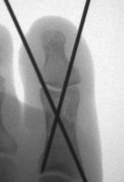
 I was able to get before and after pictures of my po' toe from the hospital, and they're so fantastic I have to share them! You can see (left) the very cracked and crunchy-looking fracture in the lower bone at the left, and then the straightened bone on the right with the pins in position. The surgeon said that he was using live x-ray imaging during the surgery to check the position of the bones and adjusted the pins as needed. Cool!
By the way, to get these pictures, all I had to do was call the imaging library at Providence (see below) and for a modest fee they gave me a CD containing the x-rays from the surgery. I don't know how long hospitals keep these images, but if you're interested in getting a copy of a scan you've had, it might be worth a call.


No comments:
Post a Comment